
Introduction: When Neck Pain Isn’t Just “Tension”
Cervical Lordosis: it’s important to understanding your neck’s natural curve and what happens when it’s lost.
Many people assume neck pain comes from stress, sleeping wrong, or spending too much time looking at a screen. And sometimes that’s true.
But for many patients, persistent neck stiffness, headaches, or shoulder tightness comes from something deeper: a change in the natural curve of the neck.
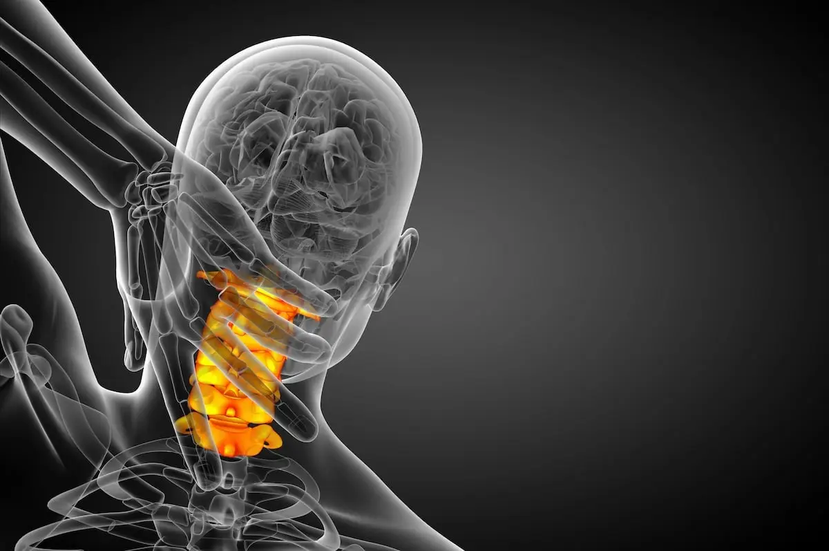
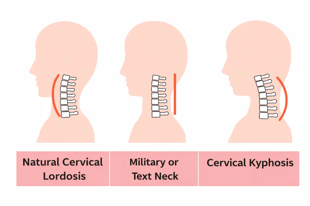
The cervical spine is designed with a gentle inward curve called cervical lordosis. This curve helps support the weight of the head, distribute mechanical forces, and keep the spine balanced.
When that curve becomes straightened, reduced, or reversed, the mechanics of the neck change — and over time this can contribute to pain, muscle fatigue, and nerve irritation.
Understanding the difference between normal cervical lordosis and loss of cervical lordosis is an important step toward relieving symptoms and protecting long-term spinal health.
In this guide we’ll explore:
-
What cervical lordosis is
-
What a normal neck curve looks like
-
Symptoms of loss of cervical lordosis
-
How doctors diagnose alignment problems
-
Exercises that may help improve comfort and posture
What Is Cervical Lordosis?
Quick Answer: Cervical lordosis refers to the natural inward curve of the cervical spine (the neck). This curve is most often described as measuring between 20 and 40 degrees on a standing side X-ray, though measured values in healthy people vary more widely than that range suggests. The curve allows the spine to support the weight of the head while maintaining balance and absorbing mechanical stress. When it becomes reduced, straightened, or reversed, it is called loss of cervical lordosis, which may contribute to neck pain, stiffness, headaches, and nerve irritation.
Understanding Normal Cervical Lordosis
The cervical spine consists of seven vertebrae (C1–C7) that support the head and allow the neck to move freely.
But the spine is not meant to be straight.
Instead, the cervical region naturally forms a gentle inward arc that works together with the curves in the thoracic and lumbar spine to keep the body balanced.
Why the Neck Needs This Curve
The human head weighs 10–12 pounds on average.
When the cervical spine maintains its natural curvature:
-
Weight is distributed evenly through the vertebrae
-
Muscles do not need to work excessively
-
Discs absorb shock efficiently
-
Nerves exit the spine without compression
This curve acts like a shock-absorbing spring, helping the spine adapt to movement, posture changes, and physical activity.
When the curve changes, the mechanical load on the neck increases significantly.
What Is the Normal Angle of Cervical Lordosis?
Quick Answer: Cervical lordosis is most commonly described as 20 to 40 degrees on a standing side X-ray, and that range is a reasonable general reference. But normal varies more than a single range suggests. Studies of people with no neck symptoms have found average values below 20 degrees in some groups, with wide variation between individuals. The measurement method also changes the number, so an angle only means something when you know how it was taken.
Patients often want a single number, and I understand why. A number feels like an answer.
But this is one of those places where the honest version is more useful than the tidy version.
What the research actually shows
The 20 to 40 degree range gets repeated almost everywhere online, and it is a fair general reference point. It is not wrong. It is just narrower and more confident than the underlying evidence supports.
When researchers have measured cervical curves in people who have no neck symptoms at all, the results vary considerably. One study of more than 300 healthy young adults found an average lordosis of roughly 17 degrees. Another normative study reported an average near 21 degrees, with a spread wide enough that both a 9 degree curve and a 33 degree curve fell within one standard deviation of average. A large review pooling data from thousands of asymptomatic individuals reached the same conclusion: cervical curvature varies substantially from person to person, even among people who feel completely fine.
So a measurement slightly outside the commonly quoted range is not automatically abnormal.
The measurement method changes the number
This part surprises most people, and it is worth understanding, because it explains why two reports on the same neck can disagree.
There is more than one accepted way to measure a cervical curve on an X-ray. The C2 to C7 Cobb angle is the most widely used. The Harrison posterior tangent method is another. Borden’s method measures the depth of the curve rather than an angle.
These methods do not produce the same value. In one study of people without neck pain, the Harrison posterior tangent method averaged about 34.5 degrees while the Cobb angle averaged about 26.8 degrees in the same group. Same necks. Different numbers. Nearly eight degrees apart.
That is not an error. It is simply what happens when you measure a curve two different ways.
What this means for your report
If your radiology report gives you a number, three things are worth knowing:
- Which method was used. Without it, the number is hard to interpret against any published range.
- How you were positioned. The X-ray should be taken standing, from the side, with your head in a relaxed natural position. Lying down changes the curve, and so does deliberately tucking or lifting the chin.
- Whether your muscles were guarding. A neck held tight by muscle spasm can photograph straighter than it normally sits.
Why I explain this to patients
Because a number without context invites the wrong conclusion in both directions.
I have met patients who were told their measurement was abnormal and spent months anxious about a value that sat well within normal variation. I have also seen the opposite, where a number landed inside the quoted range and a real problem got waved off.
The measurement is a data point. It earns its meaning when it is read alongside your symptoms and your exam, and not before.
Does A Straight Neck Always Cause Symptoms?
Quick Answer: No. Loss of cervical lordosis does not always cause pain. Imaging studies have found reduced, straightened, and even slightly reversed neck curves in people who feel completely fine and have no symptoms at all. A straight neck on an X-ray report is a finding, not automatically a diagnosis. What matters is whether that change in the curve actually explains the symptoms you are having.
Most people do not go looking for this information. They find it because a report landed in their patient portal, they read the words “loss of cervical lordosis,” and their stomach dropped a little.
If that is how you got here, I want to tell you something before anything else. Take a breath. That phrase sounds much scarier than it usually is.
Here is what I mean.
Some people have a straight neck and feel perfectly fine
Over the years, researchers have taken X-rays and MRIs of people who have no neck pain at all. Healthy people. People who were not seeking treatment for anything. When they measured the curves in those necks, they found something surprising. The curves varied a lot. Some were deep. Some were shallow. Some were nearly flat. A few were slightly reversed.
And those people felt fine.
This happens all over the body, not just the neck. Imaging is very good at showing us small changes in the spine. But not every change it shows is causing a problem. It is a little like finding a gray hair. It is real, it is there, and it tells you something about time and wear. It does not mean you are sick.
So when a radiology report says your neck curve is reduced, that sentence is describing a shape. It is not, by itself, telling you why your neck hurts.
Why this matters so much
I bring this up for two reasons, and they pull in opposite directions.
The first is fear. I have watched patients walk into my office already convinced that something is badly wrong, because they read one line on a report and then spent an evening on the internet. That fear is real, and it is exhausting, and a lot of the time it is not warranted. You deserve to know that.
The second is treatment. When a straight neck gets treated as the automatic cause of every symptom, people can end up in long, expensive care plans aimed at “restoring the curve” while the thing actually causing their pain goes unaddressed. That is a hard place to be. You feel like you are doing everything right, and you are not getting better.
Both of those outcomes come from the same mistake. It is treating a picture as if it were an answer.
So how do we tell the difference?
This is the real work of an evaluation, and it is not something an X-ray can do on its own.
When I see a patient with a straightened cervical curve, I am asking whether the shape on the film and the story the patient is telling me actually match up. That means looking at several things together:
- Where the symptoms are. Do the pain, numbness, or weakness follow a pattern that fits the levels of the spine involved? Or are they somewhere else entirely?
- What the exam shows. Reflexes, strength, sensation, and range of motion tell me things a picture cannot.
- How it started. A curve that changed after a car accident is a different conversation than one that developed slowly over ten years at a desk.
- Whether muscle guarding is involved. Sometimes a neck looks straight on film simply because the muscles were tight and splinted at the moment the X-ray was taken. The curve is not gone. It was just holding still.
- What else is on the images. Disc changes, arthritis, narrowing around the nerves, or instability may be doing far more than the curve itself.
When those pieces line up, the curve is likely part of the story. When they do not, we keep looking. Either way, you get a real answer instead of a guess.
When a straight neck does deserve attention
None of this means the finding is meaningless. It is not.
Loss of cervical lordosis matters more when it comes with symptoms that suggest the nerves or spinal cord are involved, when it appeared suddenly after an injury, when it is getting worse over time, or when it shows up alongside instability or significant degeneration. Those situations need a closer look, and sometimes they need treatment.
The point is not that a straight neck is nothing. The point is that a straight neck is a clue, not a conclusion.
You are allowed to ask which one yours is.
What Happens When the Neck Curve Is Lost?
Loss of cervical lordosis can occur gradually over time or develop after injury.
Instead of forming a natural arc, the cervical spine may appear:
-
Straightened (military or text neck)
-
Flattened
-
Reversed (kyphotic)

Common Causes of Loss of Cervical Lordosis
Several factors can contribute to changes in cervical curvature:
-
Forward head posture
-
Long hours working on computers
-
Smartphone use
-
Whiplash injuries
-
Degenerative disc disease
-
Cervical spondylosis
-
Muscle imbalance
-
Poor ergonomics
-
Trauma
One of the most common contributors today is forward head posture, often associated with modern technology use.
The Biomechanics of Posture and Neck Strain
When the head moves forward just a few inches, the mechanical load placed on the cervical spine increases dramatically.
For example:
| Head Position | Effective Weight on Neck |
|---|---|
| Neutral (0 degrees) | 10 to 12 pounds |
| 15 degrees forward | About 27 pounds |
| 30 degrees forward | About 40 pounds |
| 45 degrees forward | About 49 pounds |
| 60 degrees forward | About 60 pounds |
Figures from Hansraj KK, Surgical Technology International, 2014. These values come from a computer model of the cervical spine rather than direct measurement in patients, and the study has been debated in the literature. They are useful for illustrating how quickly load increases with forward head position, not as precise measurements of any individual’s neck.
This means that looking down at a phone or laptop for extended periods can multiply the strain placed on the neck.
Over time, the muscles that support the cervical spine may fatigue, tighten, and pull the spine into a straighter alignment, contributing to loss of cervical lordosis.
Symptoms of Loss of Cervical Lordosis
When the natural curve of the neck changes, several symptoms may appear.
Some patients experience mild stiffness, while others develop chronic discomfort.

Hansraj – 2014, Surgical Technology International
Symptoms of loss of cervical lordosis may include:
-
Chronic neck pain or stiffness
-
Tension headaches
-
Limited neck mobility
-
Shoulder and upper back tightness
-
Numbness or tingling in the arms
-
Muscle fatigue from supporting the head without proper spinal alignment
These symptoms often occur because the muscles surrounding the cervical spine must work harder to support the head when the natural curve is lost.
How Loss of Cervical Lordosis Is Diagnosed
Quick Answer: Diagnosing a change in the neck curve starts with a conversation and a physical exam, not a scan. Standing X-rays of the neck from the side are used to measure the curve itself. Flexion and extension X-rays, taken while you bend your neck forward and back, are added when there is a question about stability. MRI is reserved for situations involving nerve symptoms, neurological findings, or pain that has not improved with conservative care. Imaging supports the diagnosis. It does not make it on its own.
Patients often expect this part to be all about machines. It is not, and honestly some of the most useful information I get about a neck comes from the first ten minutes of talking, before anyone has looked at a single image.
Here is how the process usually unfolds, and just as importantly, why each piece is there.
It Starts With Your Story
I want to know when the symptoms began, what makes them better or worse, whether they stay in your neck or travel down an arm, and whether anything has changed in your hands. Are you dropping things? Fumbling with buttons or keys? Do your legs feel different when you walk?
Those last questions surprise people. They sound unrelated to a neck, but symptoms in the hands or a change in the walking pattern can point to pressure on the spinal cord itself, and if that is in the picture it changes the entire path forward. So I ask, every time, even when the answer is no.
I also want to know how this started. A curve that changed after a car accident is a very different conversation than one that developed slowly over fifteen years at a desk. Same finding on the film. Different meaning.
Physical Examination
Then I examine you.
I check how far your neck moves in each direction, and where the motion stops. I test strength in specific muscle groups, check reflexes, and test sensation along the paths individual nerves travel. I look at how you hold your head and shoulders when you are just standing there, not thinking about it.
What I am doing is building a map. If your symptoms follow the path of a particular nerve root, that tells me where to look, and if your reflexes come back unusually brisk that raises a different question entirely, one that has more to do with the spinal cord than with any single nerve. The exam narrows the field before imaging ever enters the picture, and it keeps me from chasing findings on a scan that have nothing to do with why you came in.
Standing X-Rays: Measuring the Curve
This is the study that actually shows the curve.
The important detail is that it should be taken standing, from the side, with your head in a natural resting position. Lying down changes the curve. So does deliberately tucking your chin or lifting it. Position matters more than most people realize, and a poorly positioned film can make a normal neck look flat or a flattened neck look fine.
From that image, we measure the angle of the curve. We also look at disc height, arthritic changes in the small joints, bone spurs, and how your head sits over your shoulders.
One thing worth knowing: if your muscles were tight and guarding when the picture was taken, the neck can look straighter than it usually is. The curve is not gone. It was just holding still, the way you might tense up before a flu shot, and that is one reason I do not treat a single X-ray as the final word on anything.
Flexion and Extension Films: Checking for Instability
These are the ones most patients have never heard of, and they answer a question a standard X-ray simply cannot.
A regular side X-ray is a snapshot. It shows the neck in one position, holding still, which is fine for measuring a curve but tells you nothing about what happens when the neck actually moves. Some problems only reveal themselves in motion. Flexion and extension films are two additional images, one taken while you gently bend your head forward, one while you tilt it back, and comparing them shows how the vertebrae move in relation to each other.
What I am looking for is whether any level is shifting more than it should, or whether a segment has stiffened up and stopped moving altogether.
That matters for a few reasons. Excess motion at one level can irritate nerves and cause symptoms that come and go depending on your position, which is exactly the pattern patients find so confusing to describe, and it is also the kind of thing that changes the treatment plan meaningfully. Instability is not treated the same way as a stiff, arthritic neck. If surgery ever enters the discussion, knowing which one you have is essential.
I order these when the history suggests trauma, when symptoms shift noticeably with position, when the standard films raise a question, or when we are evaluating a neck that has had surgery before.
MRI: When It Helps and When It Does Not
MRI is the study people ask for by name, and I understand why. It feels thorough.
Sometimes it is exactly the right test. But an MRI answers a specific type of question, and asking it too early can create more confusion than clarity.
An MRI is appropriate when:
- You have numbness, tingling, or weakness traveling into an arm or hand
- The exam suggests pressure on the spinal cord, including changes in your hands, balance, or walking
- Symptoms began after a significant injury
- Neck pain has not improved after a reasonable course of conservative treatment, usually around six weeks
- There are warning signs that need to be ruled out, such as unexplained weight loss, fever, night pain, or a history of cancer
An MRI usually is not the right first step when:
- The problem is straightforward neck pain and stiffness with a normal neurological exam
- Symptoms started recently and have not yet been given a chance to respond to treatment
Here is my honest reasoning, and I say this to patients often. MRI scans of healthy adults commonly show bulging discs, dried-out discs, and arthritic changes in people with no pain whatsoever, and those findings become more common with every decade of life, which means that if we scan too early we may find something real, assume it is the culprit, and start treating a picture instead of a person.
That is not caution for its own sake. It is how people end up with the wrong treatment for the right symptoms.
Other Studies You May Hear About
CT scans show bone in far more detail than an MRI does. They are useful for evaluating complex fractures, examining a prior fusion, or when an MRI cannot be performed safely because of a pacemaker or certain implants.
EMG and nerve conduction studies measure how well specific nerves are working. These help when it is unclear whether symptoms in an arm are coming from the neck or from somewhere further down, like the elbow or wrist, since carpal tunnel and a pinched nerve in the neck can feel remarkably similar to the person experiencing them.
Putting It Together
The goal is never to collect the most images. It is to answer one question clearly.
Does what we see on the film explain what you are actually feeling?
When the story, the exam, and the imaging all point the same direction, we have a diagnosis worth treating. When they disagree, that disagreement is information too, and it means we keep looking rather than settling for the first plausible answer.
You should leave a spine evaluation understanding not just what the scan showed, but what it means for you specifically. If that connection has not been made clear, it is fair to ask for it.
Cervical Lordosis vs. Kyphosis vs. Military Neck vs. Text Neck: What the Terms Actually Mean
Quick Answer: These terms get used interchangeably online, but they describe different things. Cervical lordosis is the normal inward curve of the neck. Cervical kyphosis is a curve that has reversed and now bends the wrong way. Military neck and straight neck describe a curve that has flattened out. Text neck and forward head posture describe a position your body holds, not a permanent change in the bones. Knowing which one applies to you matters, because they are not equally serious and they are not treated the same way.
If you have been reading about your neck online, you have probably run into five or six different terms that all seem to describe the same thing. Some of them do overlap. Some of them absolutely do not.
Part of the confusion is that a few of these are real medical terms, a few are nicknames, and a few started as marketing language. Here is a clear breakdown.
| Term | What it actually means | Is it normal? | How it shows up | What it usually means for you |
|---|---|---|---|---|
| Cervical lordosis | The natural inward curve of your neck, arcing gently toward the front of your body | Yes. This is the healthy, expected shape | Measured on a standing side X-ray | Nothing to fix. This is what a neck is supposed to do |
| Loss of cervical lordosis (hypolordosis) | The curve is reduced or flattened compared to what is typical | Sometimes. It is found in people with no symptoms at all | Seen on X-ray, often mentioned in a radiology report | A finding, not automatically a diagnosis. It matters only if it lines up with your symptoms |
| Military neck (straight neck) | A nickname for a neck that has flattened to nearly straight, named for a stiff, upright posture | Not a formal diagnosis. It is informal shorthand | Usually described after an X-ray | Same as loss of lordosis. The nickname makes it sound more official than it is |
| Cervical kyphosis | The curve has reversed. Instead of arcing inward, the neck bends outward | No. This is a true structural change | X-ray, sometimes with additional imaging | Deserves real evaluation, especially with nerve symptoms or if it is getting worse |
| Forward head posture | Your head sits forward of your shoulders instead of stacked over them | Common. Very common | Visible on exam, measurable on imaging | A position, not a deformity. Often improvable |
| Text neck | A popular nickname for forward head posture linked to phone and screen use | Not a medical diagnosis | Described by how you feel and how you sit | Describes a habit and its effects, not a structural condition of the spine |
The distinction that matters most
If you take one thing from this table, take this.
Text neck and forward head posture describe a position. Cervical kyphosis describes a structure.
A position is something your body is doing right now, and positions can change. A structure is the actual shape of the bones and how they sit in relation to each other. Those are very different problems, and they call for very different conversations.
Most people who search for “text neck” do not have cervical kyphosis. They have a posture pattern, some muscle fatigue, and a neck that is tired of holding a heavy head in a forward position for hours a day. That is a real problem worth addressing. It is not a spinal deformity.
Why “military neck” causes so much confusion
I hear this term from patients more than almost any other, and it is worth naming why.
“Military neck” sounds like a diagnosis. It has a definite ring to it. But it is a nickname, not a clinical classification, and no two sources define it exactly the same way. Some use it for any flattened curve. Others use it only for a completely straight one.
So when someone tells you that you have military neck, the useful follow-up question is simple. What did the measurement actually say, and does it explain what I am feeling?
Where these terms overlap in real life
To be fair, these categories are not sealed off from one another. Long-standing forward head posture may contribute over time to a flattening of the curve. A flattened curve can, in some people, progress toward kyphosis. Terms blur because the underlying conditions sometimes blur too.
But the direction of that relationship is not automatic, and it is not universal. Plenty of people hold their head forward for decades and keep a perfectly normal curve. Plenty of people with flattened curves never develop kyphosis.
The terms are useful for describing what is happening. They are not useful for predicting what will happen next. That is what an actual evaluation is for.
Exercises That May Help Improve Neck Comfort
For mild alignment changes or posture-related symptoms, targeted exercises may help improve neck support, pain and reduce muscle tension.
These exercises focus on strengthening deep neck stabilizers and correcting posture.
However, anyone experiencing severe pain, numbness, or persistent symptoms should consult a physician before beginning new exercises.
Examples of Helpful Exercises
Chin Tucks
Helps strengthen deep neck muscles and improve posture:
Sit or stand with your spine upright and gently pull your chin straight back as if making a “double chin,” keeping your eyes level. Hold for 3–5 seconds, then relax and repeat.
Shoulder Blade Retractions
Strengthens upper back muscles that support proper alignment:
Sit or stand tall and slowly squeeze your shoulder blades together as if trying to hold a pencil between them. Hold for 5 seconds, then relax and repeat.
Wall Posture Alignment
Encourages neutral spinal positioning:
Stand with your back against a wall so your heels, hips, shoulders, and head lightly touch the surface. Maintain this neutral posture for 20–30 seconds while gently drawing your chin backward.
Thoracic Mobility Exercises
Improves upper-back mobility that affects neck posture:
Sit upright and place your hands behind your head, then slowly rotate your upper back and shoulders to one side while keeping your hips facing forward. Hold briefly and repeat on the opposite side.
Gentle Cervical Extension
Helps counter forward head posture:
While sitting upright, slowly tilt your head backward to look slightly upward without forcing the motion. Hold for a few seconds, then return to a neutral position.
When Loss of Cervical Lordosis Requires Medical Treatment
Not every change in cervical curvature causes symptoms.
However, medical evaluation is recommended if you experience:
-
Persistent neck pain lasting several weeks
-
Progressive stiffness
-
Arm numbness or tingling
-
Weakness in the hands or arms
-
Pain following trauma or injury
Treatment Options May Include
Depending on the underlying cause, treatment may involve:
-
Physical therapy
-
Postural rehabilitation
-
Anti-inflammatory medications
-
Cervical traction or decompression
-
Targeted exercise programs
-
Lifestyle and ergonomic adjustments
In rare cases involving structural spinal problems or nerve compression, surgical treatment may be considered.
Dr. Steven Cyr’s Perspective on Cervical Alignment
From the perspective of Dr. Steven Cyr, Chief Medical Officer at SA Spine, loss of cervical lordosis is often a sign that the neck has been under long-term mechanical stress rather than a condition that immediately requires surgery. Changes in the natural curve of the cervical spine frequently develop over time due to posture strain, muscle imbalance, degenerative disc changes, or previous injury.
At SA Spine, the focus is first on identifying the underlying cause of symptoms and addressing it with the least invasive treatment options possible. Many patients benefit from conservative care such as physical therapy, posture rehabilitation, targeted exercise programs, and pain management strategies designed to reduce inflammation and restore better spinal mechanics.
Because the cervical spine is a complex structure involving muscles, discs, joints, and nerves, improving alignment and reducing strain often requires a multidisciplinary approach. That’s why SA Spine integrates non-surgical spine care, rehabilitation, and pain management alongside surgical expertise.
Dr. Cyr emphasizes that surgery is typically considered only after appropriate non-surgical treatments have been explored or when structural spinal problems such as nerve compression or significant instability — require surgical correction. For many patients, early evaluation and conservative treatment can relieve symptoms and help prevent more serious spinal degeneration over time.
Dr. Cyr also cautions patients against treating the X-ray as the scoreboard. A measurement is useful for understanding what is happening in the neck, but the purpose of treatment is to relieve pain, restore motion, and return people to the things they want to do. Many patients feel substantially better while their curve measurement stays essentially unchanged, and some see the number improve without feeling any different. When a treatment plan is built around changing an image rather than changing how a patient functions, it is worth asking why.
Key Takeaway
The cervical spine naturally forms a gentle inward curve called cervical lordosis, which helps support the weight of the head and maintain spinal balance. When this curve becomes straightened or reversed, patients may experience neck pain, stiffness, headaches, and nerve irritation. Improving posture, strengthening supportive muscles, and addressing underlying spinal conditions can often help restore comfort and protect long-term spinal health.
Neck Pain Shouldn’t Be Something You Just Live With
If you’re struggling with ongoing neck pain, Dr. Steven Cyr encourages patients to explore effective non-surgical solutions whenever possible before considering surgery.
A consultation at SA Spine is an opportunity to understand what’s happening in your cervical spine and discuss treatment options that may help restore comfort and function.
Schedule an appointment with the SA Spine team to get clear answers about your neck pain and the most appropriate path forward.
Resources – Related Topics To Curved Spine Conditions
- Mayo Clinic cervical spine information
- Dextroscoliosis: Symptoms, Causes, Treatment, and When to See a Specialist
FAQs
What is cervical lordosis?
Cervical lordosis is the natural inward curve of your neck, arcing gently toward the front of your body. It is a normal and healthy feature of the spine, not a condition or a problem. This curve helps support the weight of your head, spreads pressure evenly across the vertebrae and discs, and works together with the curves in your mid and lower back to keep you balanced. When people talk about cervical lordosis as something to be concerned about, they are usually referring to a change in that curve rather than the curve itself.
What is considered normal cervical lordosis?
Most sources describe a normal range of roughly 20 to 40 degrees measured on a standing side X-ray, but the honest answer is that normal varies more than that number suggests. Research on people with no neck symptoms at all has found averages well below 20 degrees in some groups, with wide variation from person to person. The measurement method matters too. The Cobb angle and the Harrison posterior tangent method can produce noticeably different numbers from the exact same X-ray, so a value only means something when you know how it was measured.
What causes loss of cervical lordosis?
There are several common causes, and often more than one is involved at the same time. Prolonged forward head posture from computer, phone, and tablet use is the one most people think of first. Others include whiplash and other neck injuries, degenerative disc disease, arthritis in the small joints of the neck, muscle imbalance or spasm, and previous neck surgery. Sometimes what looks like a flattened curve on a single X-ray is actually muscle guarding, meaning the muscles were tight and holding the neck still at the moment the picture was taken.
Can poor posture cause cervical lordosis loss?
Posture is one contributing factor, but it is not the whole story, and the research here is more nuanced than most articles admit. Studies comparing adults with neck pain to adults without it have found that people with pain tend to show more forward head posture, so there is a real association in adults. Interestingly, that same relationship has not held up consistently in adolescents. What this means practically is that posture is worth addressing, but blaming your phone for everything can cause you to miss another explanation that actually needs attention.
My X-ray says loss of cervical lordosis. Should I be worried?
Usually not, and I want to say that clearly, because this phrase frightens more people than almost anything else in a neck report. Loss of cervical lordosis is a description of a shape. It is a finding, not a diagnosis, and imaging studies have repeatedly found flattened and even slightly reversed neck curves in people who have no symptoms whatsoever. What actually matters is whether that finding lines up with what you are feeling, which takes a physical exam and a conversation about your history, not just a picture. If you have arm numbness, weakness, changes in your hands or balance, or symptoms that started after an injury, get evaluated sooner rather than later. If you feel fine and this turned up incidentally, it is worth discussing at your next visit, but it is not an emergency.
Is loss of cervical lordosis serious?
For most people, no. It becomes more significant when it appears alongside symptoms that suggest the nerves or spinal cord are involved, when it developed suddenly after trauma, when it is progressing over time, or when imaging also shows instability or significant degeneration. It is also more meaningful when the curve has fully reversed into kyphosis rather than simply flattened. The distinction between a finding that needs monitoring and one that needs treatment is exactly what a spine evaluation is designed to sort out.
Can cervical lordosis be restored?
This depends heavily on what caused the change and how long it has been present. When flattening comes from muscle tension, guarding, or postural habits, improvement is often possible with physical therapy, targeted strengthening of the deep neck flexors, and ergonomic changes. When it stems from structural changes such as disc degeneration, arthritis, or bone remodeling built up over many years, the curve itself is much less likely to shift significantly. Dr. Cyr’s position is that the goal of treatment should be reducing pain and restoring function rather than changing how an X-ray looks, and in his experience many patients improve substantially without their curve measurement changing at all.
What does a straight neck mean?
Straight neck is an informal term for a cervical spine that has lost most or all of its normal inward curve, appearing nearly vertical on a side X-ray. You may also hear it called military neck, which is a nickname rather than a formal diagnosis. Both terms mean the same thing as loss of cervical lordosis, just in more casual language. Because these nicknames sound official, they often make the finding seem more alarming and more clearly defined than it actually is.
Can cervical lordosis cause headaches?
It can be part of the picture. When the neck loses its curve, the muscles at the base of the skull and along the back of the neck often work harder to hold the head up, and sustained tension in those muscles is a well-recognized contributor to tension-type headaches and cervicogenic headaches. Irritation of the upper cervical nerves can also refer pain into the head, typically felt behind the eyes or at the base of the skull. That said, headaches have many possible causes, so persistent or worsening headaches deserve proper evaluation rather than being attributed to neck alignment by default.
How is cervical lordosis diagnosed?
Diagnosis starts with your history and a physical examination, not with imaging. A standing side X-ray of the neck, taken with your head in a natural position, is what actually shows and measures the curve. Flexion and extension X-rays, taken while you bend your head forward and back, may be added when there is a question about whether any segment is moving more than it should. MRI is generally reserved for situations involving nerve symptoms, neurological findings on exam, injury, or pain that has not improved with conservative treatment.
What exercises help cervical lordosis?
The most useful exercises focus on strengthening the deep neck flexors, improving upper back mobility, and retraining head position rather than trying to force the curve back. Chin tucks, shoulder blade retractions, wall posture alignment, and gentle thoracic rotation are the common starting points, typically done in short sets several times a day rather than in one long session. Consistency over weeks matters far more than intensity in any single session.
A note of caution: this one is not for everyone. If you have spinal stenosis, numbness or tingling in your arms, or a history of dizziness with neck movement, skip this exercise and check with your physician first.